Quick Enquiry

How Hospitals Can Evaluate 3T MRI Equipment for Better Imaging
MRI has emerged as an essential imaging technique to look inside soft tissue, organs, nerves, joints and other features of the body. Magnetic field strength is one of the key aspects that affect an MRI. It is critical for experts to understand the principle of the magnetic field strength to accurately assess the capabilities of the imaging.
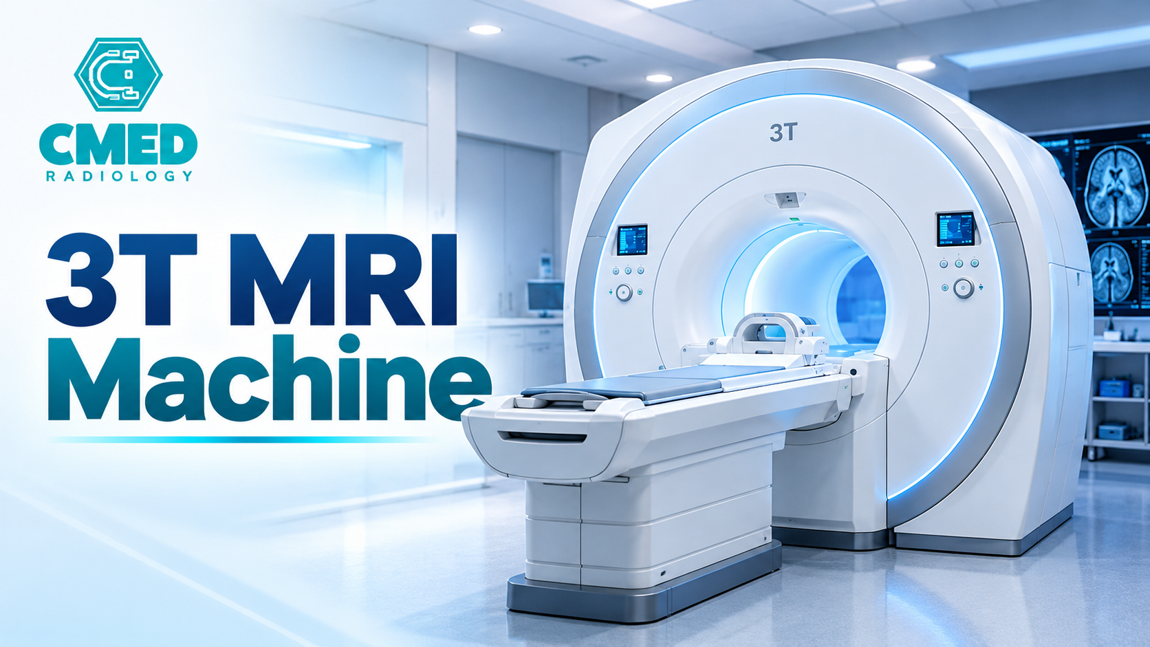
In a 3T MRI Machine, the 3 Tesla refers to the magnetic field strength of the MRI scanner. T MRI Machine is a high-field system, it is particularly suitable for use in imaging areas that benefit from an increased signal-to-noise ratio, such as studies performed in radio-isotope scan procedures.
There is more to how strong a magnetic field is than how much it pulls on metal. Information from an MRI scan is just as much about the software, coils, settings, and the person being scanned. These factors all interact to create the images. Here, we delve into the science of the strength of the magnetic field and how it impacts image quality, clinical uses and the technical factors of today's MRI systems.
What Is Magnetic Field Strength in MRI?
Magnetic Field Strength This refers to the strength of the static magnetic field which is produced by an MRI scanner. A magnetic field is necessary for the production of the signals which are used by the system to make images.
The strength of an 3T MRI Machine magnet is indicated in units called Tesla (T) which is an international unit of measurement for magnetic flux density. Most MRI equipment would generally be 1.5 Tesla or 3 Tesla, though both can provide clinically usable images, depending on the exam, scanner design and imaging protocol.
What is the Tesla Measurement?
The Tesla rating indicates the strength of the magnetic field, not the quality of the scanner as a diagnostic tool. The 3 Tesla scanner has a static magnetic field roughly twice that of the 1.5 Tesla.
Enhanced field strength also alters the response of the hydrogen protons found in the body tissue. Hydrogen protons will interact with the magnetic field and the radiofrequency energy producing signals which are received and converted into the image.
The images produced are now analyzed by radiologists to examine how normal tissue differs from abnormal tissue. 3T MRI Machine imaging, unlike conventional x-ray imaging and CT scanning, makes no use of ionizing radiation.
Why Magnetic Field Intensity Is important
Magnetic field strength influences the quantity of the signals for MRI, tissue contrast, spatial resolution of the image and imaging protocols. Higher field strength can also be exploited to produce more signal, either for greater resolution (detail in the image), or for shorter scan times.
However, the application and benefits of using a higher field will be sequence and system dependent. This is an aspect that is not a quantity in itself but rather a variable among other parameters to be taken into account.
How a 3T MRI Machine Creates Medical Imaging
To understand high-field MRI one needs to think about how the 3T MRI Machine utilises the interactions of the particles in the magnetic field to generate the images that guide our diagnoses.
1. Alignment of Hydrogen Protons
The human body is composed of water and fat both containing hydrogen. Upon locating the patient in the scanner, the main magnetic field causes the alignment of the hydrogen protons in the body to line up. This results in a net magnetisation which can be detected and is the basis of the MRI signal.
2. Use of radio frequency pulses
The 3T MRI Machine produces radiofrequency pulses at a specific frequency (i.e. A specific magnetic field strength). The RF pulses have been calculated to have the ability to alter the proton magnetisation equilibrium. As soon as the radiofrequency pulse is stopped, the protons are returned to the state of equilibrium, and these signals are picked up by the receiver coils.
3. Spatial Encoding Using Gradient Fields
A gradient field refers to the magnetic field which is built up on top of any existing magnetic field to provide information regarding which part of the body the signal originated from.
The geographic data is required for the individual image slices as well as the three-dimensional data.
4. Signal Processing and Image Reconstruction
The computer of the scanner then reads the signals that are received through the use of mathematical techniques of picture creation. The reading of the signals then can make images that show the nature of the structure of the body and the physiological disparities.
The radiologist looks at the images and thinks about what he or she knows about the patient's symptoms, medical history, and other clinical information.
A 3T MRI Machine shares the core principles of all other MRI machines, but the increased main magnetic field produces a different signal and introduces additional considerations when creating a sequence.
How Magnetic Field Strength Influences Image Quality
Image quality can be affected by magnetic field strength in a variety of ways. The extent of the effect will be determined by how the scanner is set up and how the exam is conducted.
Better Signal-to-Noise Ratio
The signal-to-noise ratio (SNR) is the amount of useful MRI signal compared to background noise. The higher the SNR, the easier it is to see the difference between anatomy and noise.
With a 3T MRI Machine, the higher signal available under similar circumstances can be exploited in various ways. Depending on the clinical requirement, imaging groups have the option of acquiring higher spatial resolution, acquiring a higher quality image or acquiring the image in a shorter scan time.
The enhancement is not the same for every body part or for each of the imaging sequences. Receiver coils, patient size, scan parameters, and reconstruction techniques all affect the final result.
Greater Potential for Spatial Resolution
The spatial resolution of a scanner relates to its ability to resolve small structures within the image that are in close proximity. High available signal allows for the use of smaller voxel sizes (volumes of an image). This can be especially useful when looking at the fine anatomy, e.g. individual regions of the brain, small joints, cartilage or nerves.
However, a higher resolution is likely to mean a decrease in the amount of signal coming from each voxel. In this case the radiologist has to decide whether to sacrifice details of the image in order to make the scan shorter and also to decrease noise.
Higher tissue contrast in Selected Examinations
3T MRI Machine contrast is variations in signal intensity between various tissues. These contrast differences are used by radiologists to separate out structural components and spot irregularities.
Tissue relaxation times and signal intensity characteristics change as the strength of the magnetic field increases. While this can be of an advantage to some studies, it can also pose problems as many artefacts also increase with field strength. Thus, the choice of sequence needs to be based on the clinical question and not on the field strength.
3T MRI Machine vs 1.5T MRI Machine
Diagnostic imaging may use either 1.5T or 3T. This will depend on workload and number of exams needed, the existing infrastructure and also the clinical needs.
This table shows general differences and not a guarantee that one system will excel over the other for every exam. A good optimiser 1.5T scanner can have extremely good diagnostic quality images, whereas a badly optimised high-field system may not reach its maximum potential. They should determine their clinical needs when selecting between the field strengths.
Clinical Applications of High-Field MRI Technology
CMED Radiology Private Limited is an additional signal generated by the 3T MRI Machine that can be used to perform a variety of imaging types. When and where to use the additional power should be based on the patient, protocol and radiology team.
Neurological Imaging
Visualisation of soft tissues and smaller structures can be visualised for brain and spinal cord scans. MRI with a high magnetic field may also allow some patients with epilepsy, multiple sclerosis, brain tumours and other neurological diseases to be scanned.
These can also be used when system factors allow - other advanced options available include functional MRI, diffusion imaging and magnetic resonance spectroscopy.
Musculoskeletal Imaging
3T MRI Machine is frequently used for joint, muscle, tendon, ligament and cartilage imaging. There are cases where higher spatial resolution may benefit a few types of exam, especially if visualization of small structures or subtle tissue changes are needed.
These may be incidental or related to trauma evaluation for sports, joint abnormalities or spinal abnormalities. The clinical benefit will depend on the imaging protocol and the interpretation.
Vascular and Cardiac Imaging
All can be measured with MRI: blood vessels, heart structure, flow, and some tissue information. However, cardiovascular imaging also requires advanced imaging protocols, breath and motion control, and a high level of experience. No cardiac study will be optimally enhanced by the higher magnetic field.
Oncology & Soft Tissue Evaluation
MRI allows clinicians to see soft-tissue abnormalities, some lesion characterisation and the extent of disease in selected cases. Apart from a little extra anatomical detail, obtained with high field imaging in a few instances, the radiologist may be able to see the relationship of abnormal tissue with adjacent structures. The image changes should always be interpreted in conjunction with other investigations.
Technical Factors That Influence MRI Performance
Receiver Coils Radiofrequency
The receiver coils are sensitive to signals from the patient's tissue. The shape of the receiver coils, the method used to increase their sensitivity, the size and shape of the receiver coils relative to the area being examined can all affect the image quality. Each coil type and system configuration are different and must be uniquely set up per high field scanner to produce a high quality scan.
Gradient System Performance
Coils inform you about the location in space that is needed to produce an image. The rate at which the gradient coils are able to turn on and off will determine the rate at which the image can be taken, the spatial resolution of the image and the type of sequences that are possible. A breakdown of the production parameters when looking at various parts of the equipment including the main magnetic field of the scanner.
Software and Reconstruction Methods
It also receives and processes the received signals and creates the final image. Utilising high-end image reconstruction techniques can be used for various purposes in image quality, workflow and specialised applications. What is pertinent to the health center is the overall software program compatibility, the series of sequences it is able to carry out, the body composition and its growth options.
Position of the Patient and Movement
Motion is an extremely prevalent cause for poor image quality and loss of diagnostic information. Correct patient positioning, fixation/immobilisation when required, good communication and optimised scan protocols will all contribute towards minimising motion artefacts. No matter how good a magnet you produce the images can be made useless by the imaging protocol or patient motion.
Safety Considerations in High-Field MRI
Unlike 3T MRI Machine does not use ionising radiation but because it uses static magnetic fields, time-varying magnetic fields and radio-frequency energy, screening processes are in place because of potential safety issues.
Screening for Implants and Metallic Objects
The patient must provide some medical information known by the MRI staff before the procedure (medical implants, previous surgeries, shrapnel, etc.) Some implants may be safe in MRI only in specific conditions, while others could be considered to be too dangerous. The team must establish what device is involved, and follow the safety rules that are relevant, rather than assuming an implant will always be safe in an MRI. All loose ferromagnetic objects in the room can be pulled by the magnetic field towards the magnet.
Radiofrequency Heating and Acoustic Noise
Radiofrequency energy can produce heat. It is the reasoning behind certain exposure limits and why the steps for monitoring the system are included in the running of the 3T MRI Machine. Another benefit of gradient switching is the loud knocking or banging noise it produces. The use of hearing protection and patient communication is another benefit seen within the MRI.
Devices and Space Needs
A high field MRI must be properly sited with a magnetic safety system, a power supply, a cooling system and trained personnel. What details are necessary may differ between scanner types or from different manufacturers. The requirement will be determined by the operating location, in conjunction with any relevant technical literature or safety requirements and local legislation. MRI safety is good equipment design, good people, good screening and good procedure.
Considerations When Evaluating MRI Equipment
All healthcare organisations thinking of buying an MRI should not take the Tesla rating at face value but instead need to review a structured assessment of each MRI system to determine whether it meets your organisation's clinical and operational needs.
Key points to consider:
Workload: what sort of examinations most frequently carried out and if any special needs for imaging.
Work flow: Review example clinical images, including the sequence images provided, for common procedures.
System configuration: Check the magnet specs, receiver coils, gradient capabilities, software, accessories.
Infrastructure: Confirm room dimensions, shielding, power, cooling and installability.
Work and service: Approximate technical support, emergency spare requirement, service record and point of care servicing.
Additional costs: Consider the costs of installation, operation, maintenance, upgrades and support of the equipment in use.
Another line of medical imaging equipment available from us used MRI units. The used MRI systems are displayed for health care providers to browse and to learn about their particulars before acquiring them.
Conclusion
The magnetic field strength is a basic component of the MRI system, affecting the type of signal generated, the possible image resolution, the tissue contrast and the scanning protocol used. A 3T MRI Machine is 1.5T stronger than a 1.5T MRI system and capable of detailed imaging when combined with appropriate coils, software, scan protocols and expert technical support.
Nevertheless, increasing the magnetic field strength does not always mean you will get a better exam, in all cases. Clinical needs, system setup, patient condition and safety procedures all influence the performance of imaging.
This information allows better information about the selection and implementation of MRI technology. Our range of medical imaging solutions will help hospitals and diagnostic centres get an idea of the available MRI options and its technicalities. The ideal 3T MRI Machine is one that provides the right mix of imaging performance at the right moment for the right application, right patient, right clinician and right use.
Frequently Asked Questions
1. What does 3 Tesla refer to in an MRI scanner?
Three Tesla is the strength of the scanner's main static magnetic field, which is roughly double the field strength of our 1.5T MRI systems.
2. Will a more powerful magnetic field always lead to higher quality MRI images?
Not necessarily. A higher field strength can give you extra signal and allow for higher detail, but there are other contributing factors such as receiver coils, protocols, software, movement and the body part being imaged.
3. What is the main advantage of a 3T MRI Machine?
The main technical benefit of the 3T MRI Machine is the higher signal available under comparable conditions. This can enable better spatial resolution, specialised advanced techniques, or shorter scan duration if the protocol can be optimised for such.
4. What is the difference between a 3T system and a 1.5T MRI scanner?
The key difference is the strength of the magnetic field. A 3T typically provides a greater amount of available signal, while a 1.5T can deliver outstanding diagnostic images with the potential to be more suitable for some scans due to variations in susceptibility effects and radiofrequency behaviour.
5. Where can healthcare providers explore MRI equipment options?
Healthcare providers can also access our portal and browse its range of medical imaging devices. They can also ask about their process needs and the MRI systems on offer.